目的 评价磷酸化细胞外信号调节激酶(p-ERK)在七氟烷致新生大鼠海马神经细胞凋亡中的作用。方法 出生7 d的健康清洁级雄性SD大鼠36只,体质量11~18 g,采用随机数字表法分为对照组(C组,n=18)和七氟烷麻醉组(S组,n=18)。C组每天同一时间吸入2 h的30%氧气;S组吸入2 h的3.4%七氟烷。2 d后处死大鼠采用Western blotting法检测活化caspase-3和p-ERK的表达。结果 与C组比较,S组活化caspase-3的表达上调,p-ERK的表达下调(P<0.05)。结论 七氟烷通过下调p-ERK的表达促进新生大鼠海马神经细胞凋亡。
七氟烷对新生大鼠海马caspase-3和磷酸化ERK表达的影响
吕淼淼 杨现会△
郑州大学第二附属医院麻醉科,河南郑州 450003
作者简介:吕淼淼,Email:miaomiao80904@126.com
【摘要】 目的 评价磷酸化细胞外信号调节激酶(p-ERK)在七氟烷致新生大鼠海马神经细胞凋亡中的作用。方法 出生7 d的健康清洁级雄性SD大鼠36只,体质量11~18 g,采用随机数字表法分为对照组(C组,n=18)和七氟烷麻醉组(S组,n=18)。C组每天同一时间吸入2 h的30%氧气;S组吸入2 h的3.4%七氟烷。2 d后处死大鼠采用Western blotting法检测活化caspase-3和p-ERK的表达。结果 与C组比较,S组活化caspase-3的表达上调,p-ERK的表达下调(P<0.05)。结论 七氟烷通过下调p-ERK的表达促进新生大鼠海马神经细胞凋亡。
【关键词】 七氟烷;细胞凋亡;海马;细胞外信号调节激酶;caspase-3;SD大鼠
【中图分类号】 R-332 【文献标识码】 A 【文章编号】 1673-5110(2019)03-0233-05 DOI:10.12083/SYSJ.2019.03.046
Effects of sevoflurane anesthesia on expression of caspase-3 and phosphorylated ERK in neonatal rat hippocampus
LYU Miaomiao,YANG Xianhui
Department of Anesthesiology,the Second Affiliated Hospital of Zhengzhou Uni>v>ersity,Zhengzhou 450003,China
【Abstract】 Objective To evaluate the role of ERK signaling pathway in sevoflurane anesthesia-caused apoptosis in hippocampal neurons in neonatal rat.Methods Thirty-two pathogen-free healthy male Sprague-Dawley 36 rats,aged 7days,weighing 11-18 g,were divided into 2 groups using a random number table:control group (group C) and sevoflurane anesthesia group(group S).In group S,the rats were exposed to 3.4% sevoflurane for 2h,while in group C the rats were exposed to 30% oxygen for 2h at the same time each day.After 2days four rats in each group were sacrificed and hippocampus were removed for determination of activated caspase-3 expression and p-ERK by Western blot.Results The level of activated caspase-3 in hippocampus was significantly higher in group S than in group C (P<0.05).Compared with group C,the expression of p-ERK was down-regulated in group S (P<0.05).Conclusion Sevoflurane anesthesia causes the neuronal apoptosis in neonatal rat hippocampus through down-regulating the expression of p-ERK in hippocampus.
【Key words】 Sevoflurane;Apoptosis;Hippocampus;Extracellular signal-regulated kinases;caspase-3;Sprague-Dawley rats
七氟烷因其起效迅速,苏醒快,作用时间短,为小儿临床麻醉常用药。动物实验表明,七氟烷可引起发育期大脑神经细胞凋亡,影响认知功能发育[1-4]。临床上也有七氟烷致小儿神经功能异常的报道[5]。研究表明,在神经细胞凋亡的早期,caspase信号通路被激活[6-7]。磷酸化细胞外信号调节激酶(p-ERK)作为一种抗凋亡蛋白,可通过调节众多基因转录的表达促进抗凋亡信号传导,发挥神经保护作用。本研究拟评价p-ERK在七氟烷致新生大鼠海马神经细胞凋亡中的作用。
1 材料与方法
1.1 实验动物分组 出生7 d的健康清洁级雄性SD大鼠36只,体质量11~18 g,购自郑州大学实验动物中心。采用随机数字表法分为2组(n=18):对照组(C组)每天同一时间吸入30%氧气2 h/次;七氟烷组(S组)吸入3.4%七氟烷+30%氧气2 h。将S组大鼠置于密闭麻醉箱中,箱底铺钠石灰,箱两侧各有一小圆孔,一侧为排气孔,并接 S/5型麻醉气体监测仪(Ohmeda Detax,美国),监测氧气,七氟烷和二氧化碳浓度;另一侧为进气孔,接七氟烷麻醉挥发罐,输入3.4%七氟烷(雅培制药有限公司,美国)和30%氧气,S组吸入3.4%七氟烷2 h,氧流量为3 L/min,麻醉过程中塑料箱下垫加温毯,温度探头插入大鼠直肠,维持肛温37 ℃,维持大鼠自主呼吸,同时翻正反射消失。对照组只吸入30%氧气2h,流量为3 L/min。
1.2 方法
1.2.1 P(O2)、P(CO2)与血糖浓度检测:于麻醉结束后15 min,每组随机取2只大鼠,腹腔注射1%戊巴比妥钠30 mg/kg麻醉后立即开胸,于左心室心尖部取血100 μL,检测动脉血P(O2)、P(CO2)与血糖浓度。
1.2.2 激活型caspase-3和p-ERK检测:于麻醉结束后15 min(T1)和麻醉结束后6 h、24 h和48 h(T2-4),每组各随机取4只大鼠,麻醉处理后断头并立即冰上取新鲜海马组织,液氮速冻,然后-80 ℃冰箱保存,采用Western blotting法检测活化caspase-3和p-ERK的表达。取冻存的海马组织,匀浆,裂解后提取蛋白,采用BCA蛋白定量法测定蛋白浓度。蛋白与5x上样缓冲液混合后,37 ℃变性30 min,采用10%分离胶和5%浓缩胶SDS-PAGE电泳,转膜,激活型caspase-3抗体(1:1 000)、β-actin抗体(1:2 000,Santa Cruz公司,美国)、磷酸化ERK抗体(1:1 000,CST公司,美国)和ERK抗体(1:1 000),4 ℃孵育过夜,1:2 000二抗室温孵育2 h,TBST洗膜5 min×3次。采用ECL(Amersham公司)化学发光检测试剂盒胶片显影。
1.3 统计学处理 采用SPSS 17.0软件进行分析,正态分布的计量资料以均数±标准差(x±s)表示,组间比较采用单因素方差分析,P<0.05为差异有统计学意义。
2 结果
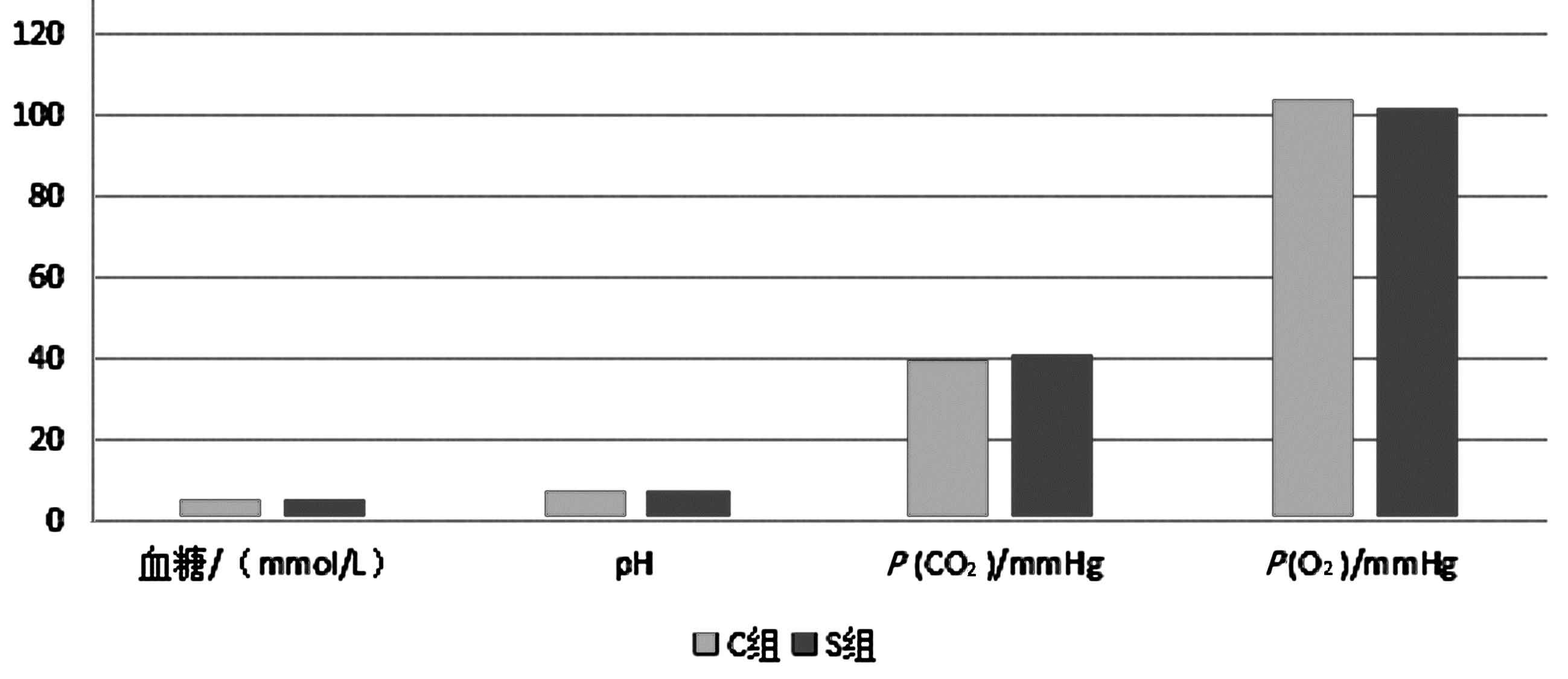
2.1 新生大鼠生理指标和血气分析检测 2组大鼠在实验过程中呼吸均匀,皮肤颜色未见明显变化,未见呼吸加深加快、呼吸暂停、呼吸停止等发生。2组pH值和血糖浓度均在正常范围。见表1、图1。
2.2 新生大鼠海马激活型caspase-3和磷酸化ERK表达的变化 与C组比较,S组T1和T2时海马活化caspase-3的表达上调(P<0.05),T1和T2时海马p-ERK的表达下调(P<0.05)。见表2。
3 讨论
新生儿期的大脑发育迅速,神经元增殖、分化等异常活跃,同时也是中枢神经系统的易感期,对内外环境变化非常敏感。研究表明[8-11],新生小鼠长时间吸入氟烷,可导致其神经细胞大量凋亡,成年后小鼠神经行为学检测也表现为异常。新生大鼠海马的快速发育期为出生后7 d[12-15],故本实验以此为研究对象。
所有吸入七氟烷的新生大鼠均在监护仪监测下进行,目的是排除呼吸和循环抑制对实验结果的影响,确保七氟烷是本实验的唯一变量指标。
Caspase又称半胱氨酸蛋白酶,其家族在细胞凋亡过程中起主要作用,分为不同的类型,但最终都需要激活caspase-3完成凋亡,在细胞凋亡中起着不可替代的作用[8,16-30]。本研究显示,新生大鼠吸入七氟烷后,海马区caspase-3被大量活化,神经细胞凋亡增多,提示七氟烷可能通过活化caspase-3诱发了新生大鼠神经细胞凋亡。动物实验显示[31-40],应用caspase-3抑制剂可明显减轻神经细胞损伤与脊髓损伤,观点支持本实验结果。
细胞外信号调节激酶(ERK)是丝裂原活化蛋白激酶(MAPK)信号转导家族成员之一。一般认为,进细胞增殖、分化与ERK信号通路密切相关。其中Ras-Raf-ERK通路是目前研究较清楚的一条信号通路[41-46]。激活的ERK参与体内诸多的生理过程,其中在神经元细胞的增殖、分化和抑制凋亡中起重要作用[47-51]。吸入七氟烷后p-ERK表达下调,凋亡神经细胞增多,提示七氟烷可能通过下调p-ERK的表达,导致新生大鼠海马神经元凋亡。因此,七氟烷致新生大鼠海马神经细胞凋亡的机制可能与下调p-ERK表达有关。
表1 2组大鼠P(O2)、P(CO2)和血糖浓度比较 (x±s)
Table 1 Comparison of P(O2),P(CO2) and blood glucose concentrations in two groups of rats (x±s)
| 组别 |
血糖(mmol/L) |
pH |
P(CO2)/mmHg |
P(O2)/mmHg |
| C组 |
4.7±0.5 |
7.38±0.03 |
39.5±1.50 |
104±10.01 |
| S组 |
5.1±0.7 |
7.37±0.02 |
41.1±1.78 |
102±11.20 |
图1 2组P(O2)、P(CO2)和血糖浓度情况
Figure 1 P(O2), P(CO2) and blood glucose concentration in two groups
表2 2组大鼠海马活化caspase-3和p-ERK比较 (x±s)
Table 2 Comparison of activated caspase-3 and p-ERK in hippocampus of 2 groups of rats (x±s)
| 指标 |
组别 |
T1 |
T2 |
T3 |
T4 |
| 活化caspase-3 |
C组 |
0.11±0.02 |
0.13±0.01 |
0.13±0.03 |
0.10±0.04 |
| |
S组 |
0.43±0.05* |
0.87±0.13* |
2.36±0.17*** |
1.12±0.08** |
| p-ERK |
C组 |
0.36±0.21 |
0.68±0.32 |
1.16±0.36 |
1.32±0.41 |
| |
S组 |
0.21±0.01* |
0.56±0.19 |
1.25±0.23 |
1.38±0.31 |
注:与C组比较,*P<0.05
4 参考文献
[1] JEVTOVIC-TODOROVIC V,HARTMAN R E,IZUMI Y,et al.Early exposure to common anesthetic agents causes widespread neurodegeneration in the developing rat brain and persistent learning deficits[J].J Neurosci,2003,23:876-882.
[2] SATOMOTO M,SATOH Y,TERUI K,et al.Neonatal exposure to sevoflurane induces abnormal social behaviors and deficits in fear conditioning in mice[J].Anesthesiology,2009,110(3):628-637.
[3] ISTAPHANOUS G K,HOWARD J,NAN X,et al.Comparison of the neuroapoptotic properties of equipotent anesthestic concentrations of deseflurane,isoflurane,or sevoflurane in neonatal mice[J].Anesthesiology,2011,114(3):578-587.
[4] 卢国林,陈小琳,宋一一,等.七氟烷吸入麻醉对新生大鼠海马神经发生和认知功能发育的影响[J].中国新药与临床杂志,2011,30(8):615-518.
[5] KEANEY A,DIVINEY D,HARTE S,et al.Postoper-ative behavioral changes following anesthesia with sevoflurane[J].Paediatr Anaesth,2004,14(10):866-870.
[6] THOMBERRY N A,LAZEBIK Y.Caspase:enemies within[J].Science,1998,281(5 381):1 312-1 316.
[7] 王建中.临床流式细胞分析[M].上海:科学技术出版社,2004:560-561.
[8] SANDERS R D,XU J,SHU Y,et al.Dexmedetomidine attenuates isoflurane-induced neu recognitive impairment in neonatal rats[J].Anesthesiology,2009,110(5):1 077-1 085.
[9] STRATMANN G,BELL J D,BICKLER P,et al.Neonatal isoflurane anesthesia causes a permanent neurocognitive deficit in rats.Program No.462.7.2006 Neuroscience.
[10] CLANCY B,FINLAY B L,DARLINGTON R B,et al.Extrapolating brain development From experimental species to humans[J].Neurotoxicology,2007,28(5):931-937.
[11] UEMURA E,lEVIN E D,BOWMAN R E,et al.Effects of halothane on synaptogenesis Meeting Planner[M].Atlanta,GA:Society for Neuroscience,2006.
[12] SATOMOTO M.Neonatal exposure to sevoflurane induces abnormal social behaviors and deficits in fear conditioning in mice[J].Anesthesiology,2009,110:628-637.
[13] LUNARDI N,ORI C,ERISIR A,JEVTOVIC-TODOROVIC V.General anesthesia causes long-lasting disturbances in the ultrastructural properties of developing synapses in young rats[J].Neurotox Res,2010,17(2):179-188.
[14] MELLON R D,SIMONE A F,RAPPAPORT B A.Use of anesthetic agents in neonates and young children[J].Anesth Analg,2007,104(3):509-520.
[15] AKER J,BLOCK R I,BIDDLE C.Anesthesia and the developing brain[J].AANA J,2015,83(2):139-147.
[16] XIE Z.The common inhalation anesthetic isoflurane induces caspase activation and increases amyloid beta-protein level in vivo[J].Ann Neurol,2008,64(6):618-627.
[17] YOUNG C.Role of caspase-3 in ethanol-induced developmental neuro-degeneration[J].Neurobiol Dis,2005,20(2):608-614.
[18] ZHANG X,XUE Z,SUN A.Subclinical concentration of sevoflurane potentiates neuronal apoptosis in the developing C57BL/6 mouse brain[J].Neurosci Lett,2008,447(2/3):109-114.
[19] COHEN G M.Caspases:the executioners of apoptosis[J].Biochem J,1997,326:1-16.
[20] WANG X,LI A J,LI W Z,et al.The effects of Xuebijing injection on apoptosis and expression of regulatory factors TNF-α、NF-κB and Caspase-3 expression in the lung tissues of acute paraquat-induced rats[J].Zhonghua Lao Dong Wei Sheng Zhi Ye Bing Za Zhi,2018,36(7):551-555.DOI:10.3760/cma.j.issn.1001-9391.2018.07.021.
[21] KO H M,JOO S H,JO J H,et al.Liver-Wrapping,Nitric Oxide-Releasing Nanofiber Downregulates Cleaved Caspase-3 and Bax Expression on Rat Hepatic Ischemia-Reperfusion Injury[J].Transplant Proc,2017,49(5):1 170-1 174.DOI:10.1016/j.transproceed.2017.03.054.
[22] LIU C,KANG Y,ZHOU X,et al.Rhizoma smilacis glabrae protects rats with gentamicin-induced kidney injury from oxidative stress-induced apoptosis by inhibiting caspase-3 activation[J].J Ethnopharmacol,2017,198:122-130.DOI:10.1016/j.jep.2016.12.034.
[23] FODOR R Ş,GEORGESCU A M,GRIGORESCU B L,et al.Caspase 3 expression and plasma level of Fas ligand as apoptosis biomarkers in inflammatory endotoxemic lung injury[J].Rom J Morphol Embryol,2016,57(3):951-957.
[24] YUAN J,REN J,WANG Y,et al.Acteoside Binds to Caspase-3 and Exerts Neuroprotection in the Rotenone Rat Model of Parkinson's Disease[J].PLoS One,2016,11(9):e0162696.DOI:10.1371/journal.pone.0162696.
[25] LOPEZ-FURELOS A,LEIRO-VIDAL J M,SALAS-SANCHEZ A A,et al.Evidence of cellular stress and caspase-3 resulting from a combined two-frequency signal in the cerebrum and cerebellum of sprague-dawley rats[J].Oncotarget,2016,7(40):64 674-64 689.DOI:10.18632/oncotarget.11753.
[26] PARK M S,JOO S H,KIM B S,et al.Remote Preconditioning on Rat Hepatic Ischemia-Reperfusion Injury Downregulated Bax and Cleaved Caspase-3 Expression[J].Transplant Proc,2016,48(4):1 247-1 250.DOI:10.1016/j.transproceed.2015.12.125.
[27] GILBERT K,GODBOUT R,ROUSSEAU G.Caspase-3 Activity in the Rat Amygdala Measured by Spectrofluorometry After Myocardial Infarction[J].J Vis Exp,2016(107):e53207.DOI:10.3791/53207.
[28] ALAN E,LIMAN N.Involution dependent changes in distribution and localization of bax,survivin,caspase-3,and calpain-1 in the rat endometrium[J].Microsc Res Tech,2016,79(4):285-297.DOI:10.1002/jemt.22629.
[29] LI Z R,TENG D H,DONG G K,et al.Expression of caspase-3 and HAX-1 after cerebral contusion in rat[J].Fa Yi Xue Za Zhi,2015,31(1):7-10;14.
[30] TALEBIZADEH N,YU Z,KRONSCHLGER M,et al.Specific spatial distribution of caspase-3 in normal lenses[J].Acta Ophthalmol,2015,93(3):289-292.DOI:10.1111/aos.12501.
[31] LIU X,ZHU X Z.Roles of p53,C-Myc,Bcl-2,Bax and Caspases in glutamate-induced neuronal apoptosis and the possible neuroprotective mechanism of basic fibroblast growth factor[J].Mol Brain Res,1999,7l(2):210-216.
[32] NAKATA N,KATO H,KOGURE K,et al.Protective effects of basic fibroblast growth factor against hippocampal neuronal damage following cerebral ischemia in the geril[J].Brain Res,1993,605:354-356.
[33] 杨新宇,杨树源,张建宁,等.急性脑创伤后神经元capase-3表达、激活及作用的实验研究[J].中华神经外科杂志,2002,18(6):375-378.
[34] MA W,YANG Y,DIAO J,et al.Sanhuangyinchi decoction pretreatment ameliorates acute hepatic failure in rats by suppressing antioxidant stress and caspase-3 expression[J].Nan Fang Yi Ke Da Xue Xue Bao,2014,34(4):482-486.
[35] YIN H Y,WEI J R,ZHANG R,et al.Effect of glutamine on caspase-3 mRNA and protein expression in the myocardium of rats with sepsis[J].Am J Med Sci,2014,348(4):315-318.DOI:10.1097/MAJ.0000000000000237.
[36] HUA P,LIU L B,LIU J L,et al.Inhibition of apoptosis by knockdown of caspase-3 with siRNA in rat bone marrow mesenchymal stem cells[J].Exp Biol Med (Maywood),2013,238(9):991-998.DOI:10.1177/1535370213497320.
[37] CETIN F,YAZIHAN N,DINCER S,et al.The effect of intracerebroventricular injection of beta amyloid peptide (1-42) on caspase-3 activity,lipid peroxidation,nitric oxide and NOS expression in young adult and aged rat brain[J].Turk Neurosurg,2013,23(2):144-150.DOI:10.5137/1019-5149.JTN.5855-12.1.
[38] LI C H,DU B,WANG P,et al.Expression of survivin and caspase-3 during the development process in rat cochlea[J].Zhonghua Er Bi Yan Hou Tou Jing Wai Ke Za Zhi,2008,43(9):686-690.
[39] SUN M,ZHAO Y,XU C.Cross-talk between calpain and caspase-3 in penumbra and core during focal cerebral ischemia-reperfusion[J].Cell Mol Neurobiol,2008,28(1):71-85.
[40] YANG B,EL NAHAS A M,THOMAS G L,et al.Caspase-3 and apoptosis in experimental chronic renal scarring[J].Kidney Int,2001,60(5):1 765-1 776.
[41] MCCUBREY J A,STEELMAN L S,CHAPPEL W H,et al.Roles of Raf/MEK/ERK pathway in cell growth,maglignant,transformation and drug resistance[J].Biochim Biophys Acta,2007,1 773(8):1 263-1 284.
[42] LIN H C,YEN C T.Differential Expression of Phosphorylated ERK and c-Fos of Limbic Cortices Activities in Response to Tactile Allodynia of Neuropathic Rats[J].Chin J Physiol,2018,61(4):240-251.DOI:10.4077/CJP.2018.BAH617.
[43] REUQUEN P,GUAJARDO-CORREA E,OROSTICA M L,et al.Prolactin gene expression in the pituitary of rats subjected to vaginocervical stimulation requires Erk-1/2 signaling[J].Reprod Biol,2017,17(4):357-362.DOI:10.1016/j.repbio.2017.10.001.
[44] SANGUEDO F V,FIGUEIREDO A M,CAREY R J,et al.ERK activation in the prefrontal cortex by acute apomorphine and apomorphine conditioned contextual stimuli[J].Pharmacol Biochem Behav,2017,159:76-83.DOI:10.1016/j.pbb.2017.07.011.
[45] TAO G,PAN L,JING R,et al.Study on Rac1/MAPK/ERK pathway mediated mechanism and role in rats with ventilator induced lung injury[J].Zhonghua Wei Zhong Bing Ji Jiu Yi Xue,2017,29(3):249-254.DOI:10.3760/cma.j.issn.2095-4352.2017.03.011.
[46] CARDOSO F D S,FRANA E F,SERRA F T,et al.Aerobic exercise reduces hippocampal ERK and p38 activation and improves memory of middle-aged rats[J].Hippocampus,2017,27(8):899-905.DOI:10.1002/hipo.22740.
[47] TANG P,DUAN C,WANG Z,et al.NPY and CGRP Inhibitor Influence on ERK Pathway and Macrophage Aggregation during Fracture Healing[J].Cell Physiol Biochem,2017,41(4):1457-1467.DOI:10.1159/000468405.
[48] MORELLO N,PLICATO O,PILUDU M A,et al.Effects of Forced Swimming Stress on ERK and Histone H3 Phosphorylation in Limbic Areas of Roman High-and Low-Avoidance Rats[J].PLoS One,2017,12(1):e0170093.DOI:10.1371/journal.pone.0170093.
[49] DOU M Y,WU H,ZHU H J,et al.Remifentanil preconditioning protects rat cardiomyocytes against hypoxia-reoxygenation injury via δ-opioid receptor mediated activation of PI3K/Akt and ERK pathways[J].Eur J Pharmacol,2016,789:395-401.DOI:10.1016/j.ejphar.2016.08.002.
[50] RAMREZ D,SABA J,CARNIGLIA L,et al.Melanocortin 4 receptor activates ERK-cFos pathway to increase brain-derived neurotrophic factor expression in rat astrocytes and hypothalamus[J].Mol Cell Endocrinol,2015,411:28-37.DOI:10.1016/j.mce.2015.04.008.
[51] YAP J J,CHARTOFF E H,HOLLY E N,et al.Social defeat stress-induced sensitization and escalated cocaine self-administration:the role of ERK signaling in the rat ventral tegmental area[J].Psychopharmacology (Berl),2015,232(9):1555-1569.DOI:10.1007/s00213-014-3796-7.
(收稿2019-01-15)
本文责编:夏保军
本文引用信息:吕淼淼,杨现会.七氟烷对新生大鼠海马caspase-3和磷酸化ERK表达的影响[J].中国实用神经疾病杂志,2019,22(3):233-237.DOI:10.12083/SYSJ.2019.03.046
Reference information:LYU Miaomiao,YANG Xianhui.Effects of sevoflurane anesthesia on expression of caspase-3 and phosphorylated ERK in neonatal rat hippocampus[J].Chinese Journal of Practical Nervous Diseases,2019,22(3):233-237.DOI:10.12083/SYSJ.2019.03.046
